Bladder tumours can be reduced by 90% using nanorobots
Bladder cancer has one of the highest incidence rates in the world, and is also the fourth most common tumour in men. Although it does not have a high mortality rate, almost half of bladder tumours recur within 5 years, requiring continuous monitoring of the patient, with frequent hospital visits and the need for repeat treatment. For all these reasons, bladder cancer is one of the most expensive cancers to cure.
- A mind-blowing journey inside the bladder
- Years of work and a spin-off
- Technological innovation in microscopy to locate nanorobots
Current treatments involving the administration of drugs directly into the bladder have shown good survival rates, but low therapeutic efficacy. A promising alternative is the use of nanoparticles, capable of delivering the therapeutic agent directly to the tumour. Particularly noteworthy are nanorobots, nanoparticles with the ability to self-propel inside the body.
Now, a study published in the prestigious journal Nature Nanotechnology shows how a research team has been able to reduce the size of bladder tumours in mice by 90% by administering a single dose of urea-powered nanorobots.
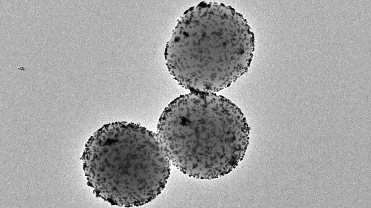
These tiny nanomachines consist of a porous sphere of silica. On their surface, they incorporate various components with specific functions. One of these is the enzyme urease, a protein that reacts with urea, which is present in urine, making the nanoparticle capable of propelling itself. Another key component is radioactive iodine, a radioisotope commonly used for localised treatment of tumours.
The work, led by the Institute for Bioengineering of Catalonia (IBEC) and CIC biomaGUNE and developed in collaboration with the Institute for Research in Biomedicine (IRB Barcelona) and the Autonomous University of Barcelona (UAB), opens the door to new treatments for bladder cancer that reduce hospitalisation time, which would imply lower costs and greater comfort for the patient.
"With a single dose we see a 90% decrease in tumour volume. It is much more efficient, taking into account that patients with this type of tumour usually go to hospital between 6 and 14 times. With this type of treatment we would increase efficiency, reducing hospitalisation time and the cost of treatment," explains Samuel Sánchez, ICREA research professor at IBEC and leader of the study.
The next step, which the team is already working on, is to study whether these tumours reappear after treatment.


A mind-blowing journey inside the bladder
In previous research, the scientists confirmed that the self-propulsion ability of the nanorobots allowed them to reach all the walls of the bladder. This is an advantage over the current procedure, where once the treatment is administered directly into the bladder, the patient has to change position every half hour to get the drug to reach all the walls.
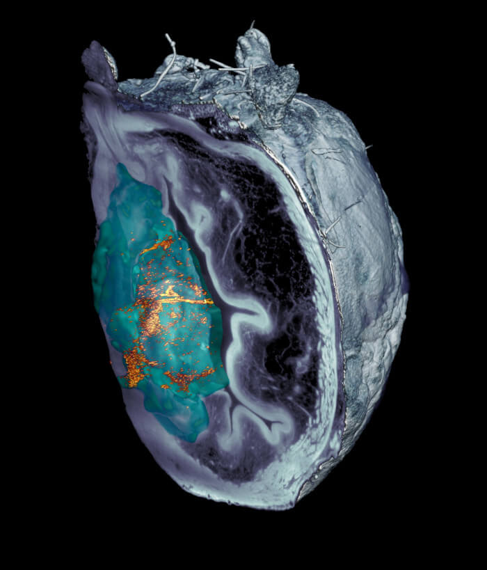
The new work goes further by demonstrating not only the mobility of the nanoparticles in the bladder, but also their specific accumulation in the tumour. This was possible thanks to different techniques, including medical positron emission tomography (PET) images of the mice, as well as microscopy images of the excised tissues after completion of the study. The latter were acquired using a fluorescence microscopy system developed specifically for this project at IRB Barcelona. The system makes it possible to observe the entire bladder, scanning the different layers of the organ and then obtaining a 3D reconstruction.
"The innovative optical system we developed allowed us to cancel out the light reflected by the tumour itself and thus identify and locate the nanoparticles throughout the organ, without prior marking, at an unprecedented resolution. We saw that the nanorobots not only reached the tumour, but also managed to access its interior, thus favouring the action of the radiopharmaceutical," explains Julien Colombelli, leader of the Advanced Digital Microscopy scientific platform at IRB Barcelona. Deciphering why nanorobots are able to access the interior of the tumour was a challenge. Nanorobots do not contain specific antibodies to recognise the tumour, and normally, tumour tissue is more rigid than healthy tissue.
"However, we observed that these nanorobots have the ability to break down the extracellular matrix of the tumour by locally increasing the pH through a self-propulsive chemical reaction. This phenomenon could favour greater tumour penetration and proved beneficial in achieving preferential accumulation in the tumour," explains Meritxell Serra Casablancas, co-first author of the study and IBEC researcher.
The scientists concluded that the nanorobots collide with the urothelium as if it were a wall, but in the tumour, being more spongy, they penetrate and accumulate inside it. A key factor is the mobility of the nanobots, which increases the likelihood that they will reach the tumour.
Moreover, according to Jordi Llop, researcher at CIC biomaGUNE and co-leader of the study, "The localised administration of the nanorobots carrying the radioisotope reduces the probability of generating adverse effects, and the high accumulation in the tumour tissue favours the radiotherapeutic effect".
"The results of this study open the door to the use of other radioisotopes with a greater capacity to induce a therapeutic effect, but whose use is restricted when radiopharmaceuticals must be administered systemically," adds Cristina Simó, co-first author of the study.
Years of work and a spin-off
The study brings together the results of more than three years of collaborative work between several institutions. Part of the data derives from the doctoral theses of Meritxell Serra and Ana Hortelao, both researchers in IBEC's Smart Nano biodevices group, led by Sánchez. Also from the thesis of Cristina Simó, co-first author of the study, who carried out her pre-doctoral research in the Radiochemistry and Nuclear Imaging Laboratory led by Jordi Llop at CIC biomaGUNE. In addition, the group at the Universitat Autònoma de Barcelona led by Esther Julián has experience in the animal model of the disease. In addition, the project has received funding from the European Research Council (ERC) and the "La Caixa" Foundation.
The technology on which these nanorobots are based, which Samuel Sánchez and his team have been working on for more than seven years, has recently been patented and is the basis of Nanobots Therapeutics, an IBEC and ICREA spin-off created in January 2023.
The company, founded by Sánchez, represents a bridge between research and clinical application: "Obtaining good funding for the spin-off is crucial to be able to continue developing this technology and, if all goes well, for it to reach the market and society. In June, only 5 months after the creation of Nanobots Tx, we successfully closed the first round of funding, and we are excited about the future," says Sánchez.
Technological innovation in microscopy to locate nanorobots
Working with nanorobots has been a major scientific challenge in bioimaging techniques for the visualisation of these elements in tissues and the tumour itself. The most common non-invasive techniques used in the clinical setting - such as PET - do not have the resolution needed to locate these very small particles at a microscopic level. For this reason, the Scientific Microscopy Platform at IRB Barcelona used a microscopy technique that involves the use of a laser light sheet to illuminate the samples and obtain three-dimensional images by scattering light when it hits tissues and particles. Observing that the tumour itself scattered some of the light, generating interference, they developed a new technique based on polarised light that cancels out all the scattering from the tissue and cells of the tumour itself. This made it possible to visualise and locate the nanorobots without the need to have previously marked them with molecular techniques.